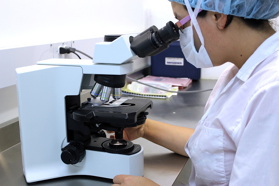
Microscope Image
'Electron microscope images of diatoms. Diatoms are a major group of algae, and are among the most common types of phytoplankton.' 'I could only exhale when looking at this digital colourisation of a scanning electron microscope image of a DIATOM' 'Colourised scanning electron microscope images of diatoms by Dr Paul Hargreaves and Faye Darling.
Microscope Images at Different Magnifications Do you need some examples of images at different magnifications under a microscope? The different images below were taken with two different kinds of microscopes. The images of Paulownia wood, hair and frog's blood were taken with a high power, also known as compound microscope using a Nikon Coolpix camera and a. The compound microscope typically has three or four different magnifications - 40x, 100x, 400x and sometimes 1000x. • At 40x magnification you will be able to see 5mm.
• At 100x magnification you will be able to see 2mm. • At 400x magnification you will be able to see 0.45mm, or 450 microns. • At 1000x magnification you will be able to see 0.180mm, or 180 microns. The images taken of the sunflower with the moth pupa were taken with a low power or stereo microscope. A stereo microscope is a good instrument for viewing insects, coins, leaves, anything you might be able to hold in the palm of your hand, but need to see more detail on the item.
Condition: New. Basic statistics by b l agarwal pdf free.
The images of the moth were taken with an Olympus camera using an. Images captured with a high power microscope and a Nikon Coolpix 5400 digital camera (5.1 mega pixels).
Pictures obtained from digital microscope are both entertaining and educational. Unfortunately, to receive a bigger view you often need to combine, align and realign microscope photographs of different parts of the sample. Not a tool complicated task, of course, but neither is it pleasant. PhotoStitcher provides a nifty and much faster way to merge multiple microscope images into a single picture. Read along to learn how to make a perfect stitching with this excitingly simple tool.
Step 1: Upload all your microscope images to the program Run PhotoStitcher and click the [+] button on the toolbar. Select all images you want to stitch together. They can be overlapping, non-aligned and such – that doesn’t matter with PhotoStitcher.
Alternatively, you can just drag and drop all images to the window. Step 2: Select wrap surface to 'Plane' Click the Stitch button on the toolbar.
* Improved keyboard queue handling slightly. * Minor config screen fixes. Pcsx2 0.9.8 standalone installer.
But before the images start merging together, you should select the mode for them. For microscope pictures select 'Scans' in the Properties dialog. Step 3: Start stitching microscope images!

Now just click OK and wait for the program to process all images. PhotoStitcher automatically aligns images and accurately matches edge to edge and quickly produces continuous picture from multiple source microscope photos.